
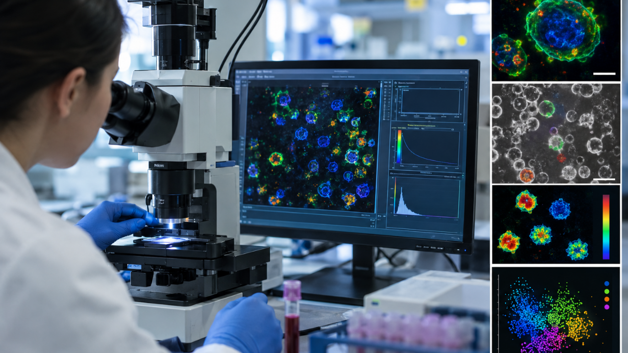

When discussing immune cell imaging, traditional methodologies have heavily relied on exogenous chemical dyes and fluorescent markers to illuminate cellular states under a microscope. However, a revolutionary scientific breakthrough has altered this paradigm entirely. Researchers have successfully developed a novel technique that captures and analyzes the natural light emitted from cellular molecules, bypassing the need for invasive dyes completely.
Table of Contents
Understanding the Dye-Free Breakthrough
For decades, visualizing the inner workings of our immune system required modifying cells with various chemical agents. While these methods advanced medical science significantly, they also introduced potential artifacts, altered cellular metabolism, and limited the scope of real-time observation. The newly unveiled immune cell imaging technique changes this by tapping into the intrinsic optical properties of living cells.
By examining thousands of individual white blood cells extracted from routine blood draws, scientists achieved an astonishing milestone. The system identified activated immune cells versus resting ones with an accuracy rate reaching nearly 94 percent, and it accomplished this comprehensive analysis within just two hours.
The Power of Single-Cell Metabolism
At the heart of this innovation lies the detailed evaluation of cellular metabolism at a single-cell level. Every living cell constantly produces and consumes energy, leaving a unique metabolic signature. By capturing the microscopic shifts in energy production, the technology can instantly tell whether an immune cell is dormant or actively fighting a threat.
How Autofluorescence Imaging Works
Cells naturally emit a faint glow when exposed to specific light wavelengths, a phenomenon known as autofluorescence. Molecules like NADH and flavins, which play critical roles in cellular energy metabolism, naturally fluoresce. By measuring these specific light emissions without adding external chemicals, researchers gain an unfiltered look into the cell’s physiological state.

Key Advantages Over Traditional Staining
- Non-Invasive: No chemical dyes or genetic alterations required, preserving natural cell behavior.
- Speed: Generates accurate diagnostic profiles within a rapid two-hour window.
- Precision: Achieves a 94 percent accuracy rate in distinguishing activation states.
- Scalability: Easily applied to ordinary, small-volume blood samples.
Implications for Medical Diagnostics
The practical applications of this technology are vast and transformative. Traditional diagnostic workflows often involve lengthy incubations and complex panel setups to evaluate immune responses. With this streamlined approach, clinicians could soon monitor patient immune health dynamically, detecting infections, autoimmune flare-ups, or vaccine responses much earlier than previously possible.
Future Directions in Immunology
As researchers continue to refine this optical analysis platform, integration with artificial intelligence and machine learning models is underway. Automated pattern recognition will likely push the accuracy even higher while reducing processing times further. This synergy between advanced optics and data science heralds a new era for personalized medicine and immunology research.
Conclusion
The introduction of this dye-free immune cell imaging technique marks a monumental leap forward in biomedical science. By providing a rapid, highly accurate, and non-destructive window into cellular metabolism, researchers can now study immune responses with unprecedented fidelity. As this technology transitions from the research laboratory to clinical settings, it holds the profound potential to redefine how we diagnose, monitor, and treat a myriad of health conditions.